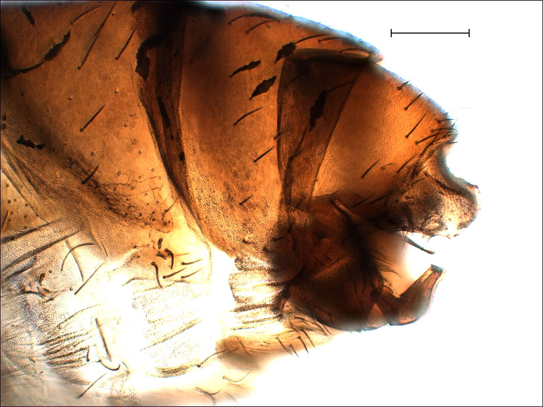
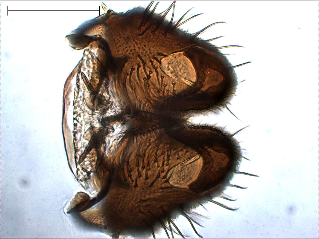
On the side and ventral views of the terminalia below, we don't see
very much, because too much stray light is degrading the resolution.
So we first pull out the ninth segment and cut the membrane with the
scalpel.
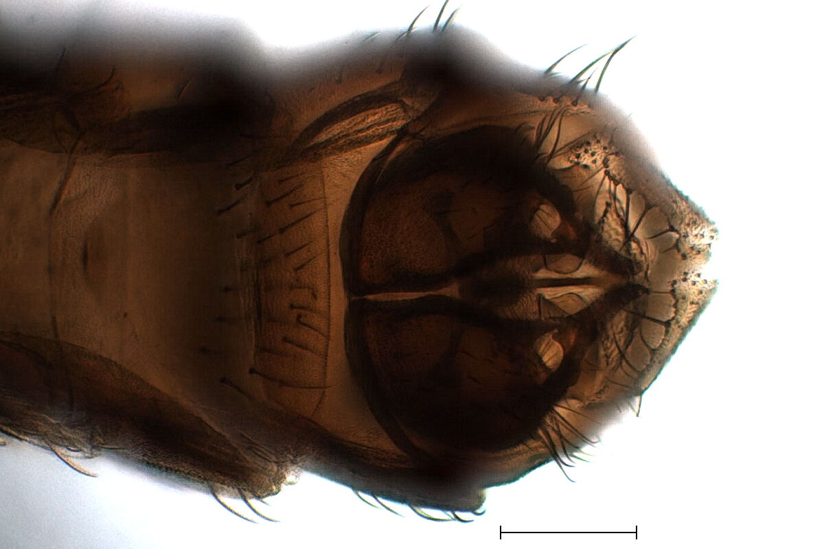
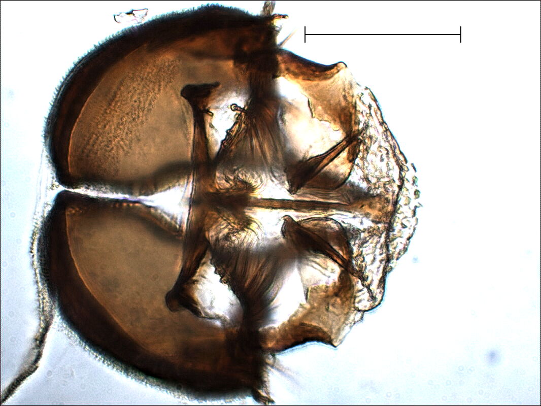
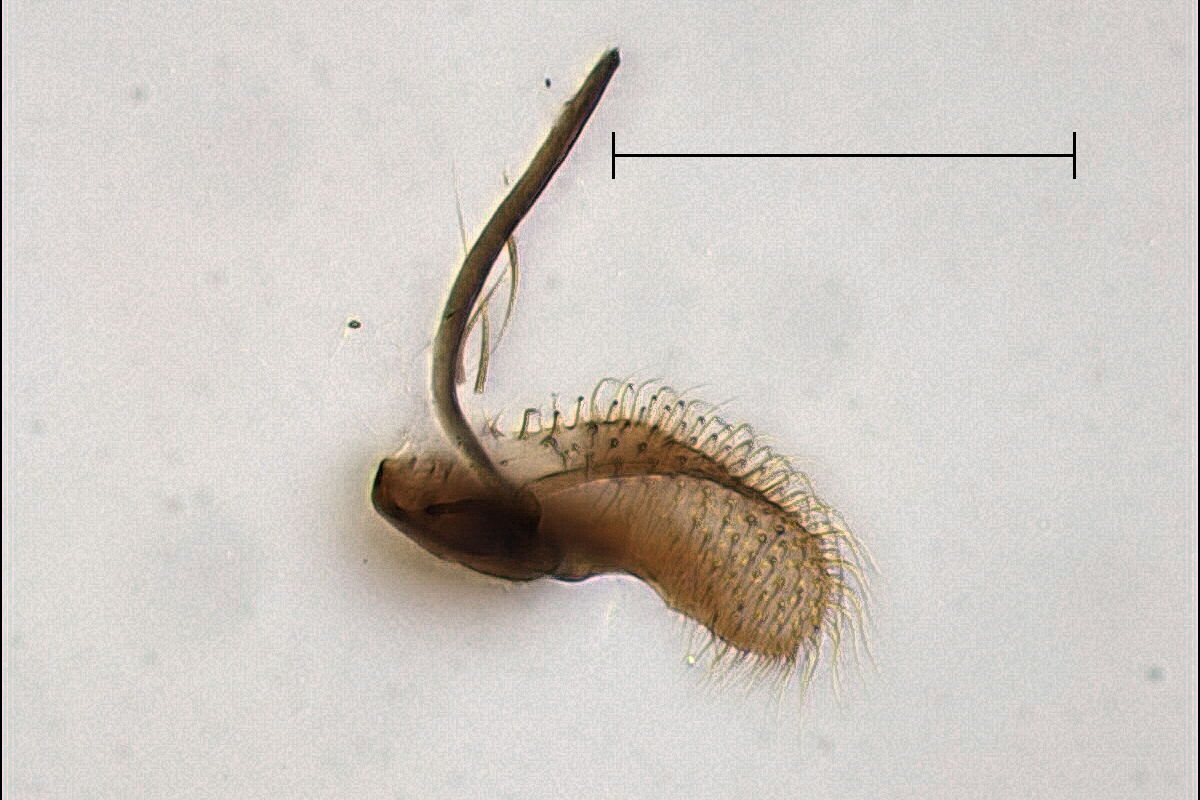
In the caudal view below we get a better idea of gonocoxites and
gynostyli, and, if we know it's there, we can even distinguish the
gonosternum. We see that there is only a membranous connection
between the gonocoxites and internal parts and the rest. We cut this
membrane, first ventrally, bend out the coxites, cut the dorsal part
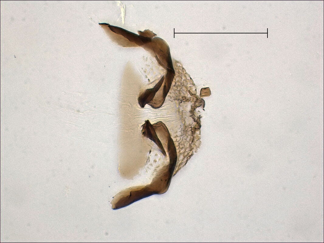
of the membrane, and pull out the coxites. On the right side below
you see the separated 9th tergite with cerci and what Rubzov calls
the tenth tergite in caudal view.
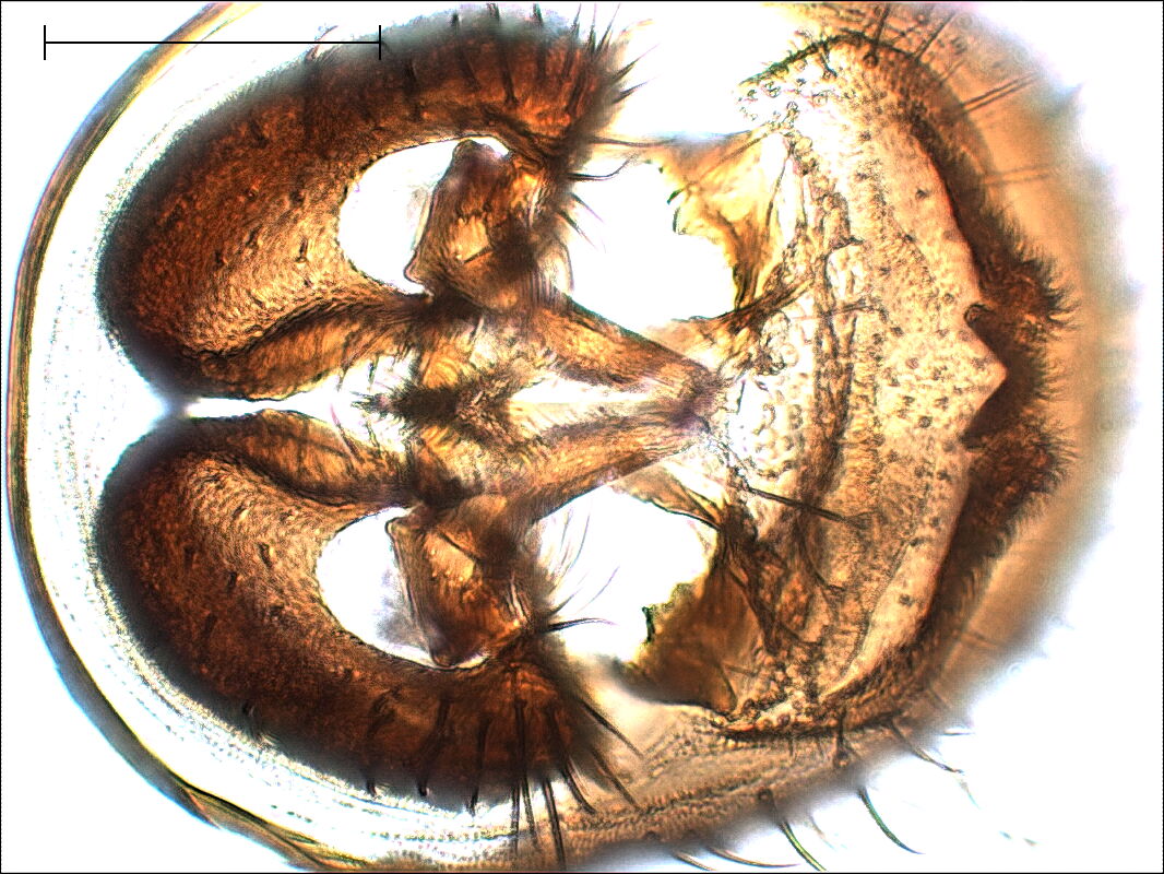
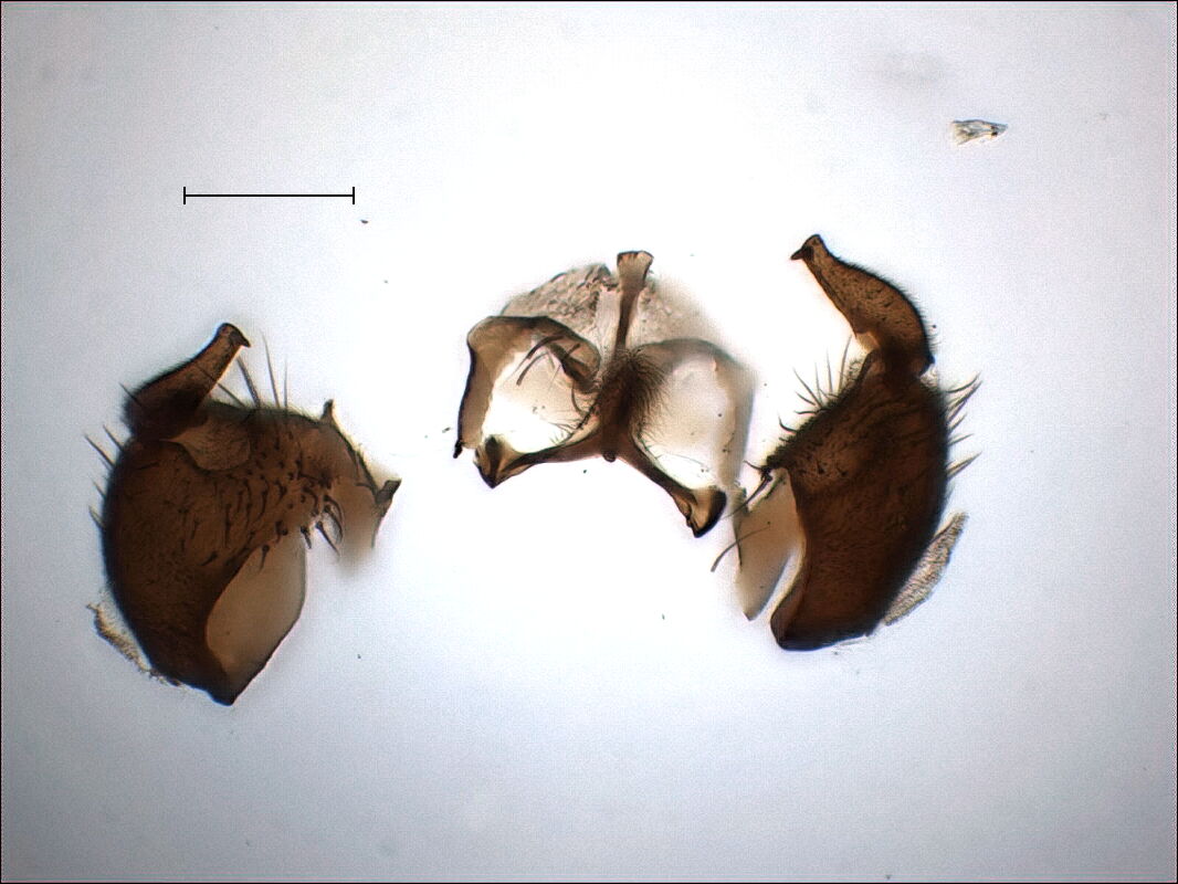
Now we can see a bit more of the gonosternum between the coxites
(dorsal view on the left), and in the anterior (inside) view on the
right we can already see the parameres. As the coxites don't share a
sclerotised connection, they can be pulled apart. We insert a blunt
pin into one of them and pull away the other one, possibly cutting
the membrane and the slightly sclerotised connection of the base of
the parameres with the coxites.
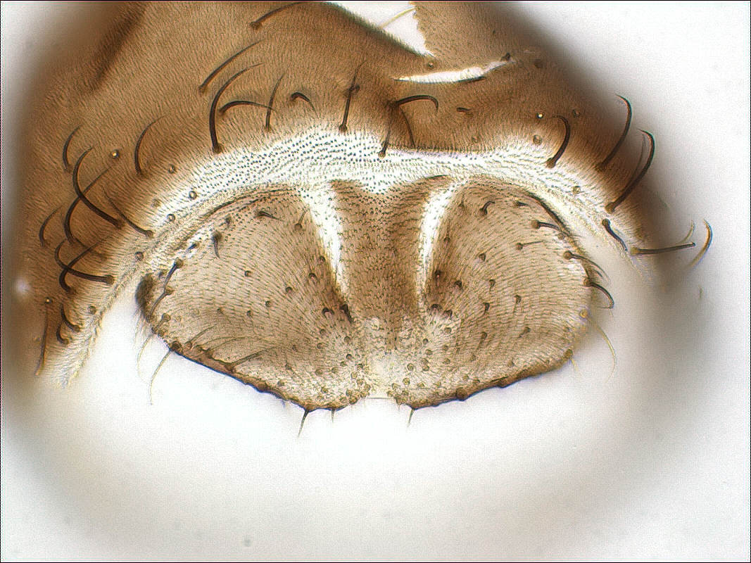
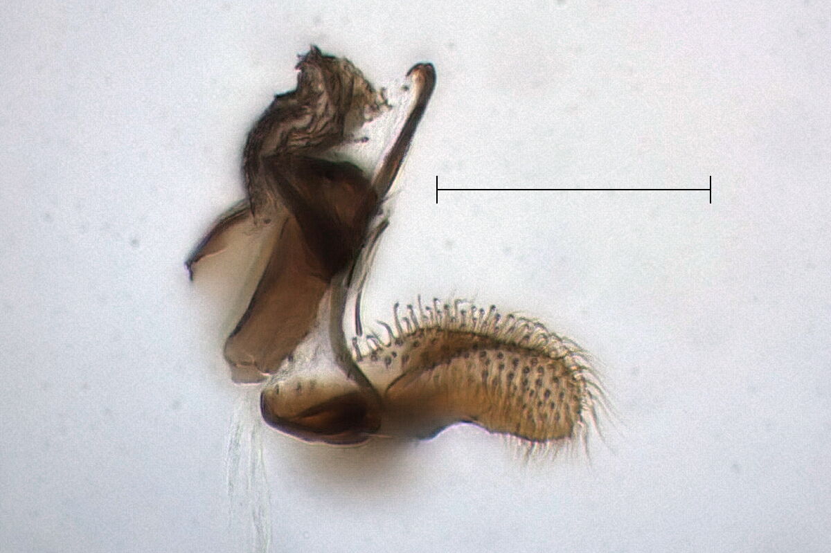
Now we have a much better view on the internal parts and can take a
first side view of parameres and gonosternum. Again these two share
only a membranous connection and can be separated.
Finally we try to get the best possible resolution by embedding.