2. Example: Helina latitarsis |
Back |
Top |
|
|
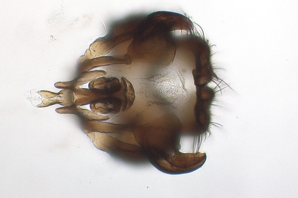
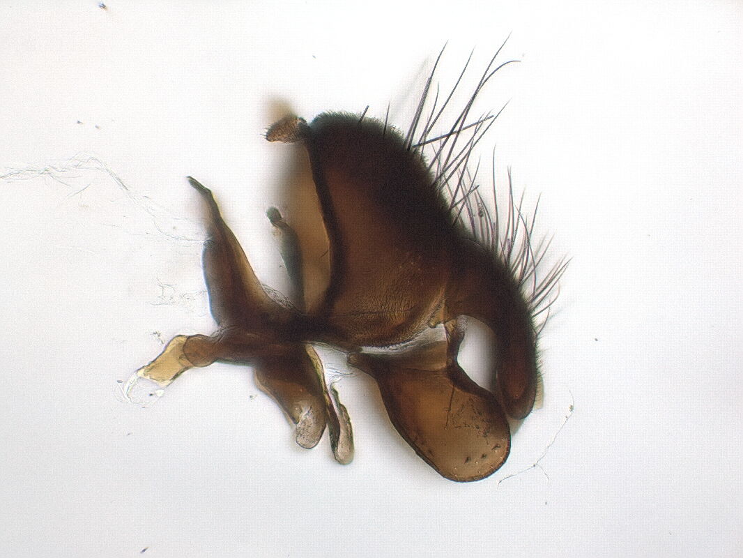
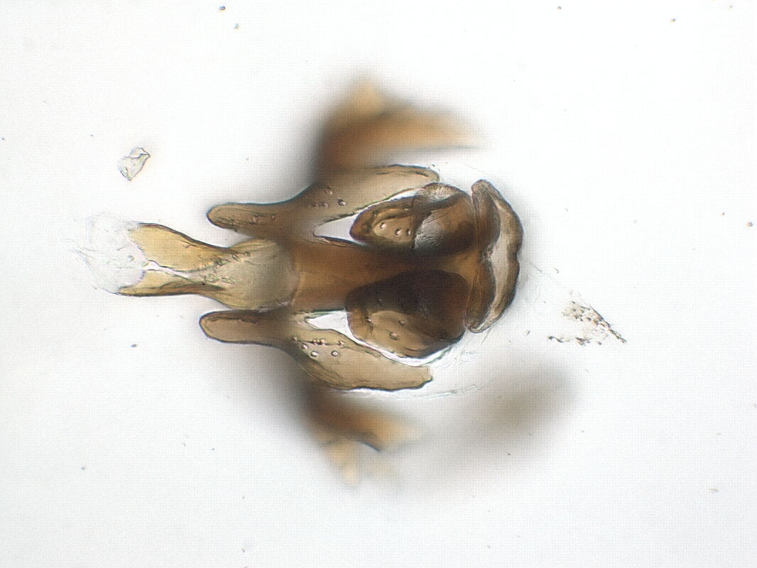
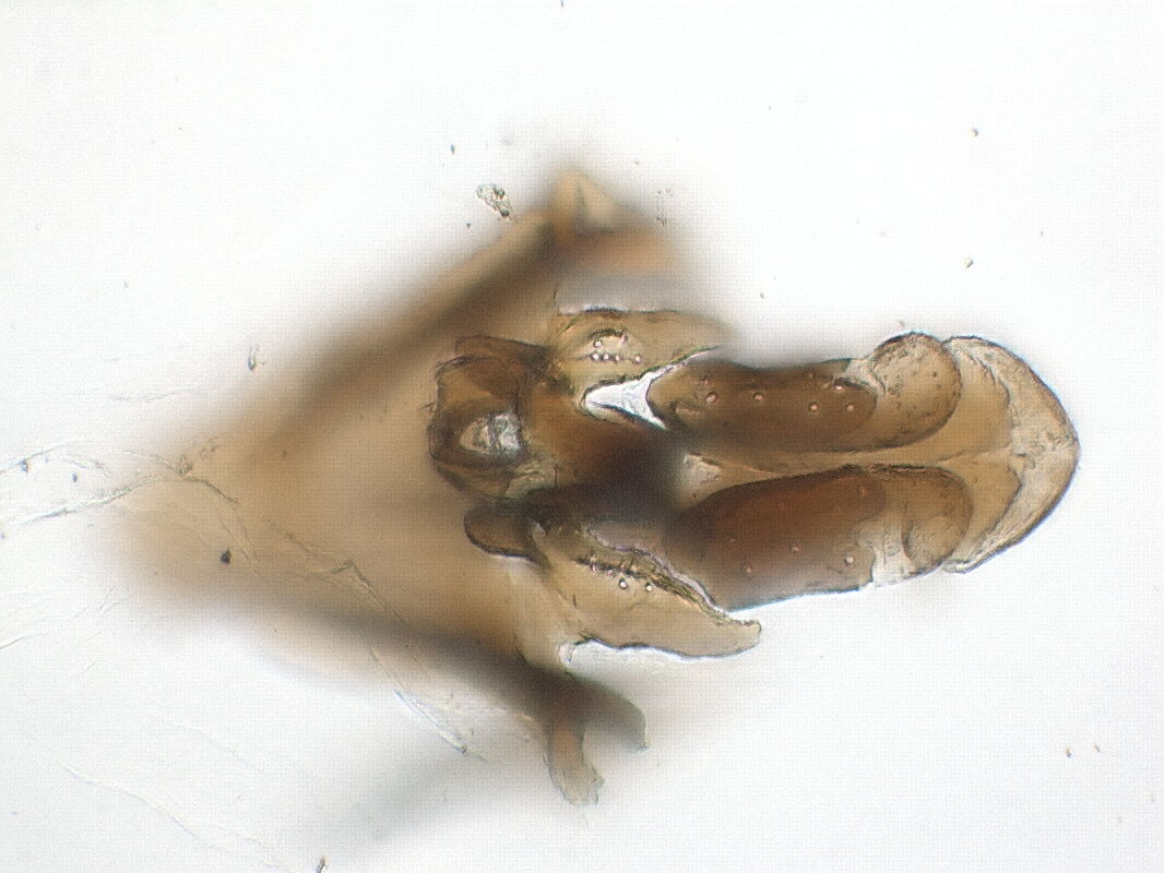
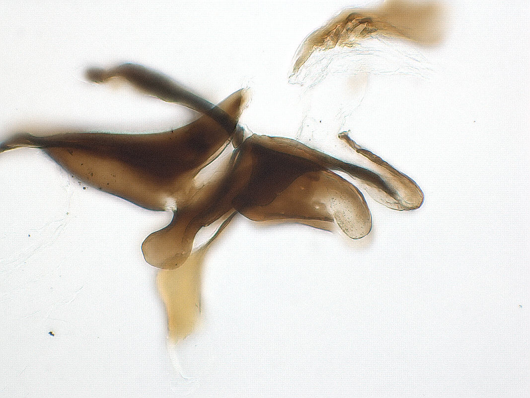
The hypandrial arms both end posteriorly in a fork, visible in the image to the left above and below the pregonites. The outer arms are connected to the middle of the anterior part of the epandrium, and the inner arms are connected via the bacilliform sclerites to the bases of the surstyli. The first step is to pierce into the membrane between the surstyli, holding the epandrium to the bottom with a blunt pin from inside. Especially in Muscidae and Anthomyiidae, the connection between bacilliform sclerites and surstyli is much more rigid, than the connection between surstyli and epandrium. If we try to separate this junction, often rather the surstyli are separated from the epandrium, and we have to show them separately, so we loose the natural orientation of the parts relative to each other. Often it is wiser to relinquish the bacilliform sclerites and break them close to the hypandrium together with the junction to the epandrium. To do this, keep the blunt pin inserted dorsally, and press down the scalpel on the pin. Now we can show the hypandrium from all sides (caudally, ventrally, and from the side), and possibly continue dissection.
 |
 |
 |