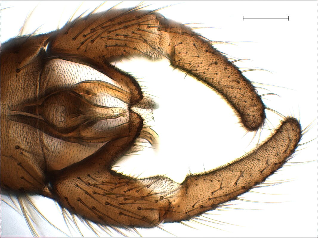
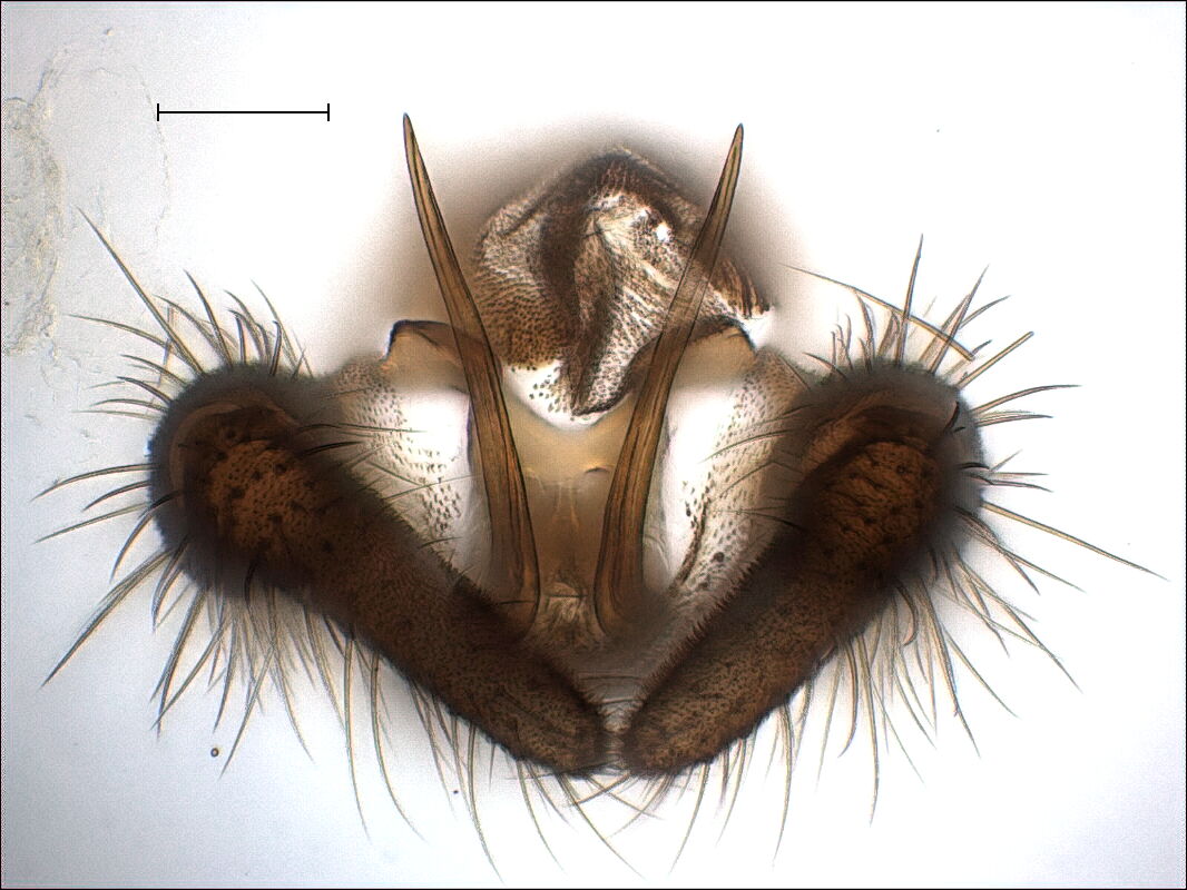
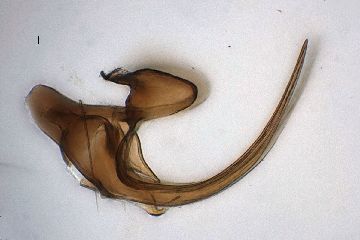
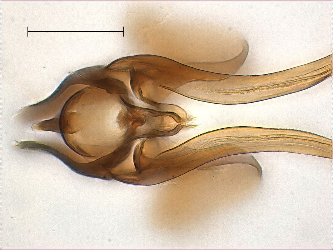
3. Example: Trichocera saltator |
Back |
Top |
|
|
We first insert a blunt pin into the open anterior side, press
down the ninth tergite to the bottom, pierce the acute scalpel
into the membrane from behind, and separate it from the rest. Now
we insert the acute scalpel from behind between the dorsal side
arm of the aedagal complex and the gonocoxite and break the
sclerotized junction. Having done that on both sides, we can pull
out the aedeagal complex and show it in dorsal, side, and ventral
view.
 |
 |
 |