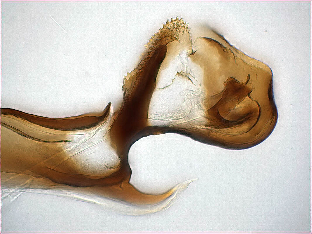
Below you see the distiphallus of Scathophaga lutaria. The side view
on the left side is taken in artificial Canada Balsam, with the best
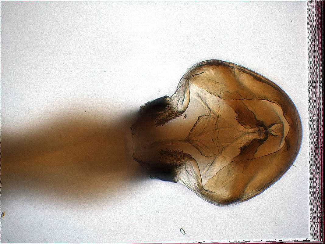
resolution possible. On the right side you see a ventro-caudal view,
showing the internal structures. On the right and bottom margin, you
can clearly see how the alignment algorithm tracked the rotation of
the object, while I was taking the series of ~30 exposures.
In this case it is important to use a video (USB, microscope) camera
and not an SLR body, because the series must be taken as fast as
possible, say with one exposure per second. With one hand on the
fine drive of the microscope and the other one on the mouse, this is
quite easy.