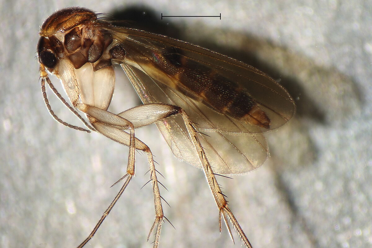
Before starting dissection we take a photo of the whole body, to
show coloration and size ratio. We hold the sixth segment, the last
"normal" one, with Dumont forceps and cut off the terminalia right
before it.
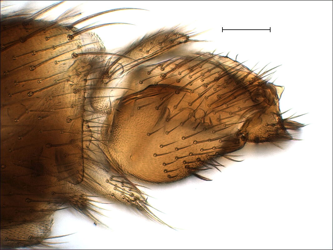
After clearing, we insert a blunt pin from the left (left photo
below), press it sideways to the bottom, pierce the acute scalpel
into the mebrane right before the ninth segment, and pull untile we
can see the reduced 7th and 8th segments. The only sclerotized link
of the ninth segment to the rest is a small ventral fork. Try to
separate the ninth segment by piercing with the scalpel exactly
between fork and the ninth segment. Remove the membranous dorsal
connection. Now we take a caudal view to get a first glimpse inside,
with the dorsal lobe of the gonstylus above and ventral one below.
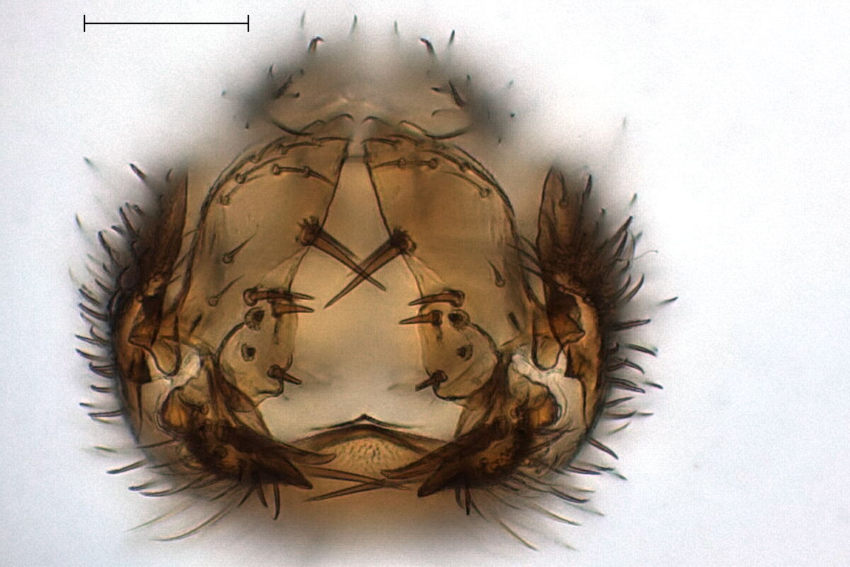
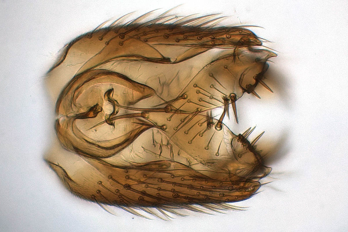
In the dorsal view on the left below we see the small ninth tergite
(left) with the cerci, both obstructing the view on the aedeagus.
The problem is that the middle part of the ninth tergite is often
weaker, than the membraneous connection to the right and left
gonocoxites. So we insert the blunt pin into the anterior side (from
the left) between aedeagus and gonocoxa and bend away the cerci.
Then we crack the membrane on both sides and pull the ninth tergite
away.
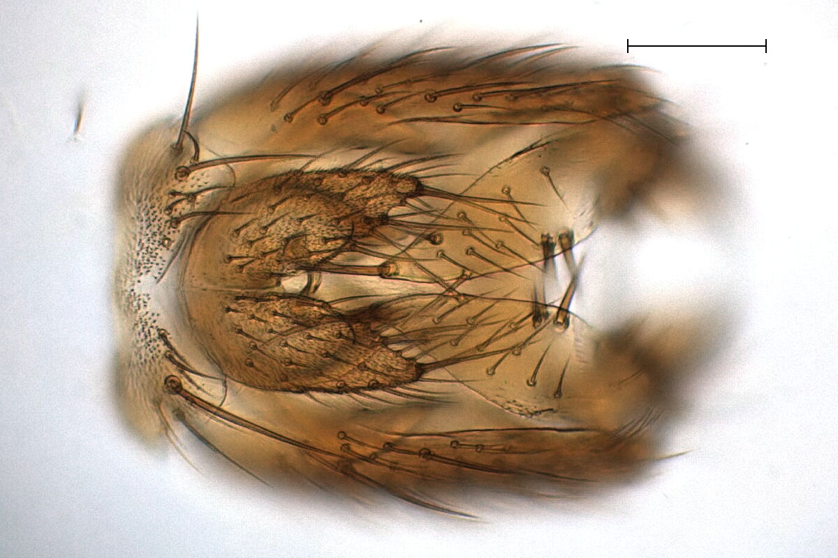
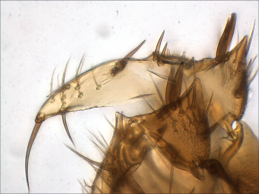
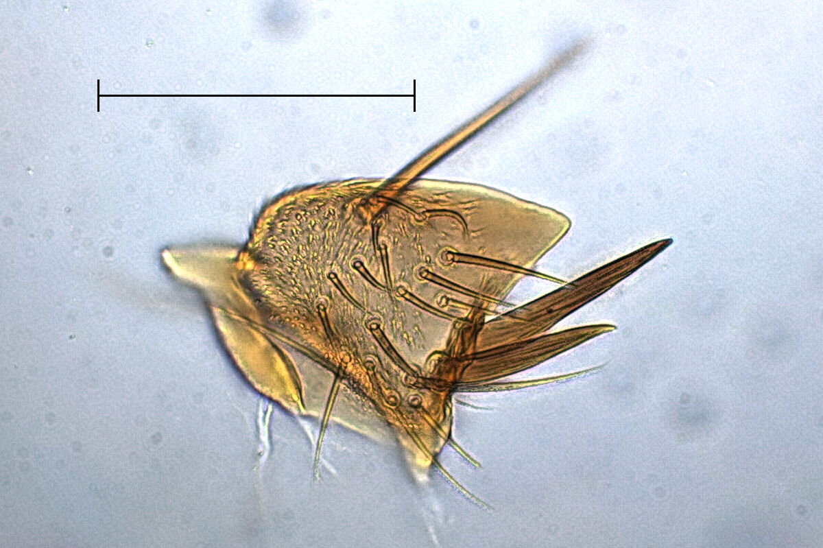
Inserting the blunt pin again between aedeagus and gonocoxite, we
press down the hypopygium to the bottom and carefully bend out the
right gonostylus. This is the only place where I use the word
"careful" in the examples, because you must watch out where you can
touch the gonostylus without loosing bristles. In the left photo
below we see the left ventral lobe in front and the whole bent out
gonostylus behing. Now find a place between right gonostylus and
gonocoxa, where you can pierce in the acute scalpel. Repeat that
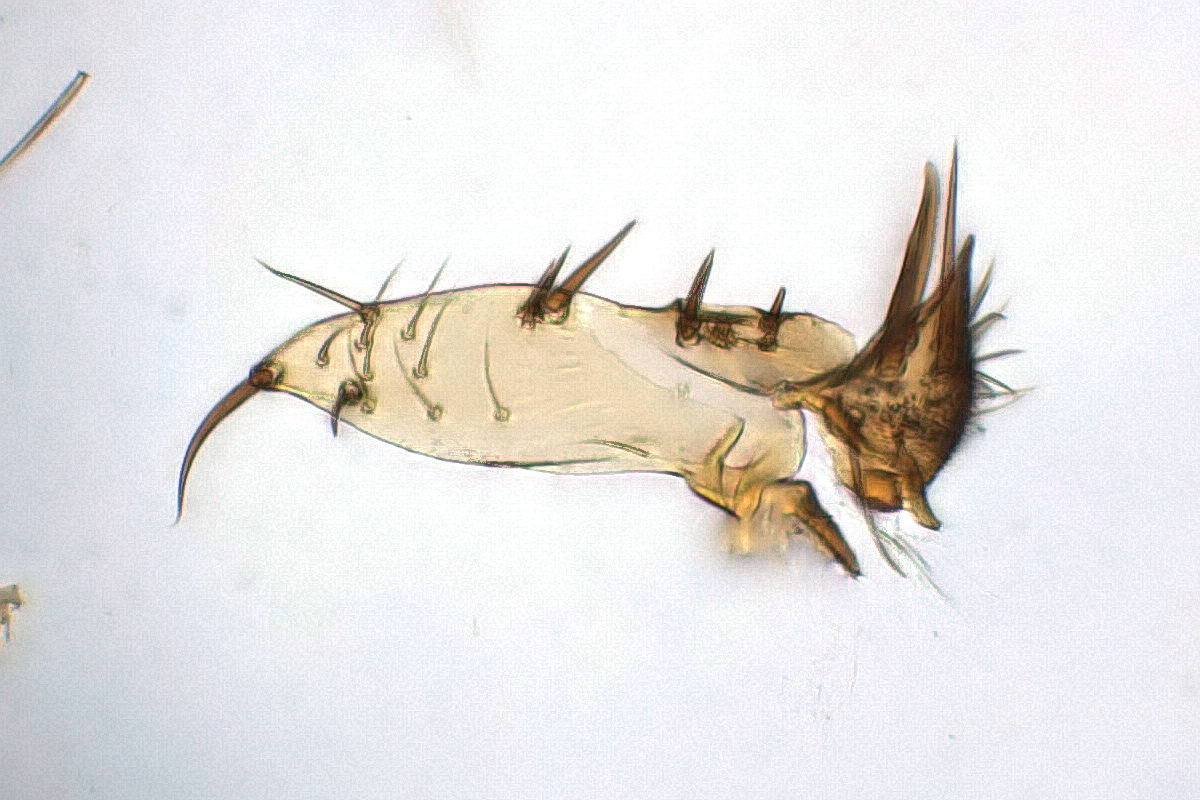
several times until you can pull it away. Now we could embed the
gonostylus for better resolution, but the natural orientation
between dorsal and ventral lobe is quite unlucky, that we better
separate them. So we use the highest magnification of the binocular,
orient the gonostylus with the scalpel and pierce it down on the
joint between them, watching for bristles, which mustn't be touched.
For such small parts I use only the right hand, holding it with the
left one against trembling.
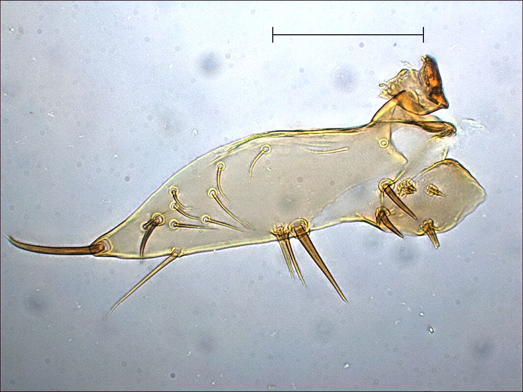
Finally we can easily embed the separate lobes, and see clearly
which of the bristles on the dorsal lobe are actually bristles and
which ones are bunches of thin hairs.