Comparison of the Resolution Achieved in Glycerol and Malinol |
Back |
Top |
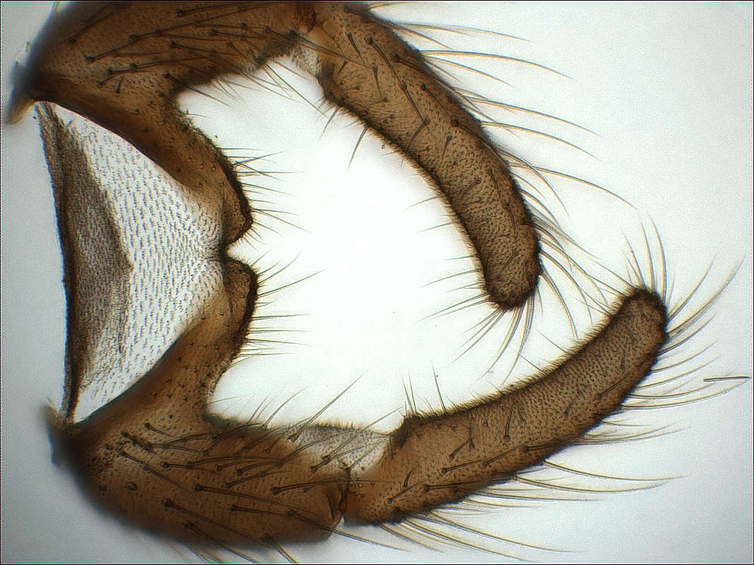
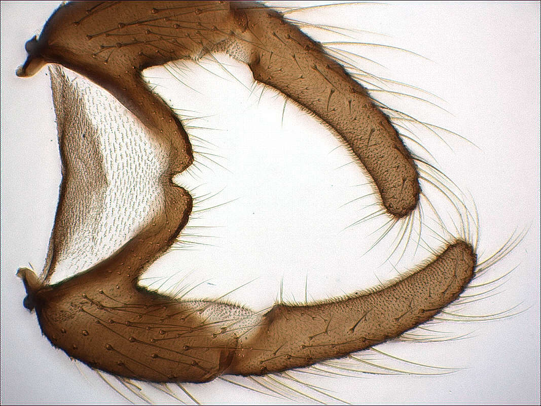
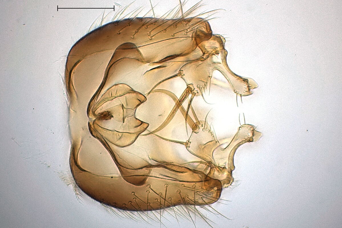
The first example shows the ventral view of the terminalia of a
Trichocera saltator, aedeagal complex and ninth tergite removed.
The main difference is, that the hairs, especially on the
gonocoxites, appear too thick, which is simply due to the worse
resolution in glycerol.
|
|
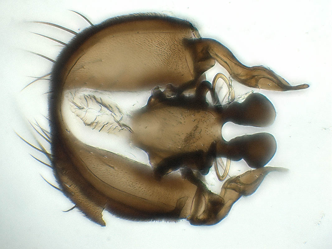
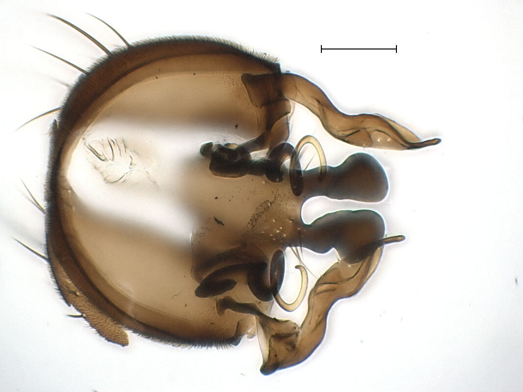
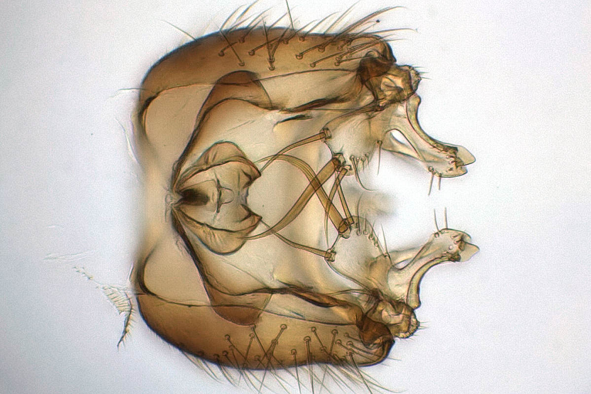
The next example shows an interior view of the epandrium of a
Fannia parva. In the Malinol photo we get a much better idea of
how the spiracle shaped bacilliform sclerites are connected to the
surstyli. It almost looks like the hinge of a door.
 |
 |
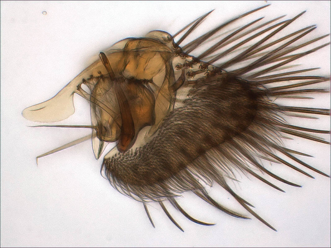
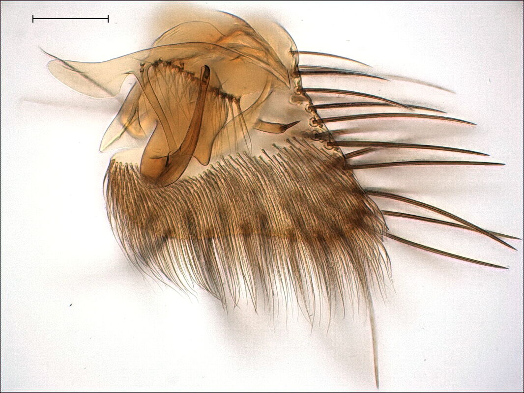
Here we see the right gonites of a Copromyza similis, postgonite
is on the left side. The small sensillae with a very fine hair in
the middle are almost invisible in glycerol.
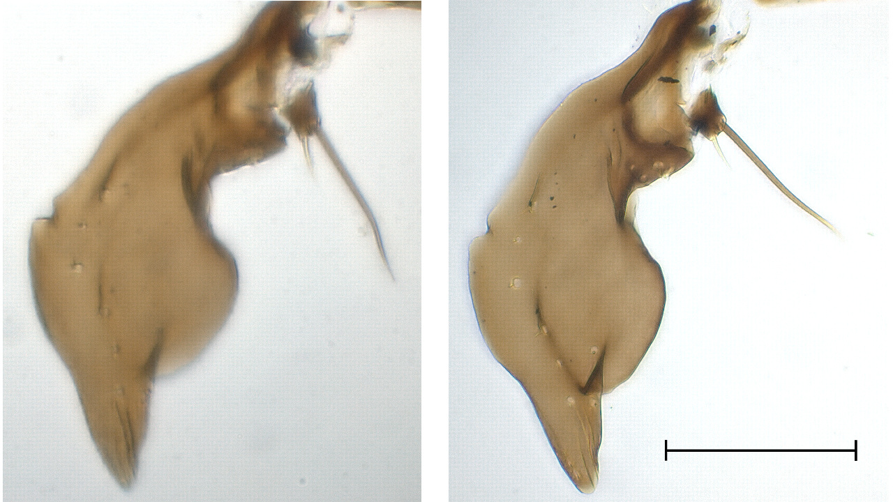 |
This is the right gonostylus of a Exechia festiva. The Malinol
photo makes it much easier to disentangle the many little lobes,
hairs, and bristles attached to the inner side of the gonostylus.
 |
 |
And finally, to keep the pros and cons in balance, an example
where use of Malinol is again questionable, the dorsal view of the
hypopygium of a Mycetophila alea, ninth tergite removed.
 |
 |